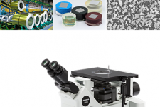
Inverted Metallurgical Microscope GX53

Overview
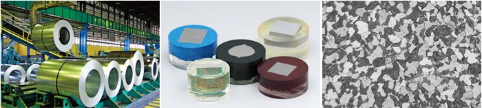
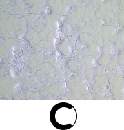
Quickly Analyze Thick, Large Sample Materials
Designed for use in the steel, automotive, electronics, and other manufacturing industries, the GX53 microscope delivers crisp images that can be difficult to capture using conventional microscopy observation methods. When combined with PRECiV image analysis software, the microscope streamlines the inspection process from observation to image analysis and reporting.
Fast Inspections, Advanced Functionality
Quickly observe, measure, and analyze metallurgical structures.
Advanced Analysis Tools
1. Combined observation methods produce exceptional images 2. Easily create panoramic images 3. Create all-in-focus images 4. Capture both bright and dark areas |
Optimized for Material Science
1. Software designed for materials science 2. Metallurgical analysis that complies with industrial standards |
User Friendly
Even novice operators can comfortably make observations, analyze results, and create reports.
1. Easily restore microscope settings 2. User guidance helps simplify advanced analysis 3. Efficient report generation |
Advanced Imaging Technology
Our proven optics and imaging technology deliver clear images and reliable results.
1. Reliable optical performance: wavefront aberration control 2. Clear images: image shading correction 3. Consistent color temperature: high-intensity white LED illumination 4. Precise measurements: auto calibration | 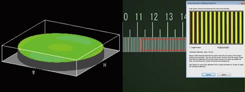 |
Modular
Choose the components you need for your application.
1. Build your system your way: fully customizable system with a variety of optional components | 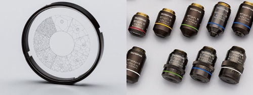 |
Observation
Advanced Analysis Tools
The GX53 microscope’s various observation capabilities provide clear, sharp images, so you can reliably detect defects in your samples. PRECiV image analysis software's new illumination techniques and image acquisition options give you more choices for evaluating your samples and documenting your findings.
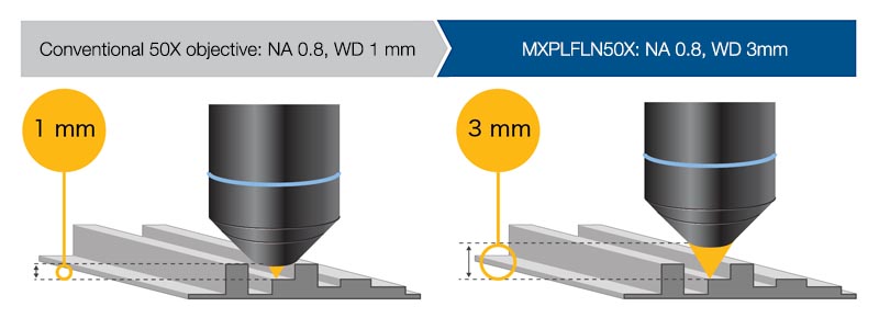
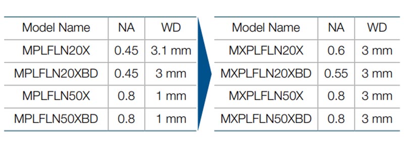
Combined high numerical aperture and long working distance
Objective lenses are crucial to a microscope’s performance. The new MXPLFLN objectives add depth to the MPLFLN series for epi-illumination imaging by maximizing numerical aperture and working distance at the same time. Higher resolutions at 20X and 50X magnifications typically mean shorter working distances, which forces the sample or objective to be retracted during objective exchange. In many cases, the MXPLFLN series’ 3 mm working distance eliminates this problem, enabling faster inspections with less chance of the objective hitting the sample.
|
|
Learn More about MXPLFLN Objectives>>
The Invisible Becomes Visible: MIX Technology
MIX technology combines darkfield with another observation method, such as brightfield or polarization, to enable you to view samples that are difficult to see with conventional microscopes. The circular LED illuminator has a directional darkfield function where one or more quadrants are illuminated at a given time, reducing a sample’s halation to better visualize surface texture.
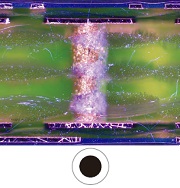
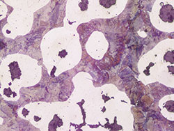
Cross-section of a printed circuit board
|
|
|
|
| ||
Brightfield | Darkfield | MIX: Brightfield + Darkfield |

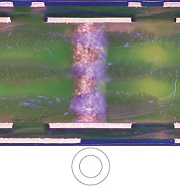
Stainless steel
|
|
|
|
| ||
Brightfield | Darkfield quadrant | MIX: Brightfield + Darkfield quadrant |
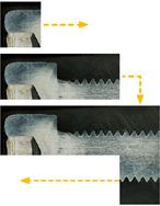
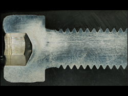
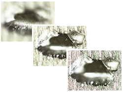
Easily Create Panoramic Images: Instant MIA
With multiple image alignment (MIA), you can stitch images together simply by moving the XY knobs on the manual stage—a motorized stage is optional. PRECiV software uses pattern recognition to generate a panoramic image, making it ideal for inspecting carburizing and metal flow conditions.
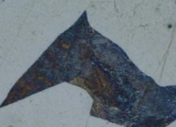
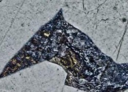
Metal flow of a bolt
|
|
|
|
| ||
Adjust the stage position using the XY knob. | The full condition of metal flow can be seen. |
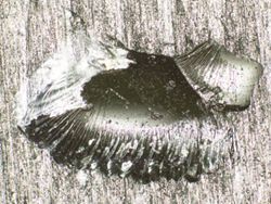
Create All-in-Focus Images: EFI
PRECiV software’s extended focus imaging (EFI) function captures images of samples whose height extends beyond the depth of focus. EFI stacks these images together to create a single all-in-focus image of the sample. Even when analyzing a cross-section sample with an uneven surface, EFI creates fully-focused images.
EFI works with either a manual or motorized Z-axis and creates a height map to visualize structures.
Resin parts
|
|
|
|
| ||
Adjust the objective’s height with the focusing handle. | EFI automatically captures and stacks multiple images to create a single, in-focus image of the sample. | Fully focused image is created. |
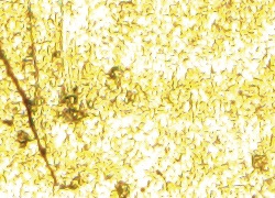
Capture Both Bright and Dark Areas Using HDR
Using advanced image processing, high dynamic range (HDR) adjusts for differences in brightness within an image to reduce glare. It also helps boost the contrast in low-contrast images. HDR can be used to observe minute structures in electric devices and identify metallic grain boundaries.
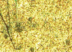
Gold plate
|
|
| ||
Some areas have glare. | Both dark and bright areas are clearly exposed using HDR. |
Chromium diffusion coating
|
|
| ||
Low contrast and unclear. | Enhanced contrast with HDR. |
Applications
There are just a few examples of what can be achieved using different observation methods.
Polished sample of AlSi (Brightfield / Darkfield)
|
|
| ||
Brightfield | Darkfield |
Brightfield: a common observation method to observe reflected light from a sample by illuminating it straight on. Darkfield: observe scattered or diffracted light from a sample, so imperfections, such as minute scratches or flaws, clearly stand out.
Spheroidal graphite cast iron (Brightfield / DIC)
|
|
| ||
Brightfield | DIC observation |
Differential interference contrast (DIC): an observation technique where the height of a sample is visible as a relief, similar to a 3D image with improved contrast; it is ideal for inspections of samples that have very minute height differences, including metallurgical structures and minerals.
Aluminum alloy (Brightfield / Polarized light)
|
|
| ||
Brightfield | Polarized light observation |
Polarized light: a technique that highlights a material’s texture and crystal condition to view metallurgical structures, such as the growth pattern of graphite on nodular cast iron and minerals.
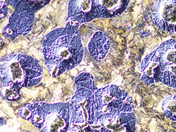
Electric device (Brightfield / MIX observation)
|
|
| ||
Brightfield | MIX: Brightfield + Darkfield |


MIX observation: combines brightfield and darkfield to show a sample’s color and structure.
The above MIX observation image clearly reproduces the device’s color and texture as well as the condition of the adhesive layer.
Analysis
PRECiV Software – Optimized for Materials Science
Together, the GX53 microscope and PRECiV software support metallurgical analysis methods that comply with different industrial standards. With step-by-step operator guidance, users can analyze their samples quickly and easily.
> Click here for details about PRECiV
Particle Analysis – Count and Measure Solution
The Count and Measure solution uses advanced threshold methods to reliably separate objects, such as particles and scratches, from the background. More than 50 different object measurement and classification parameters are available including shape, size, position, and pixel properties.
|
|
|
|
|
Conventional software | Etched steel microstructure | PRECiV |


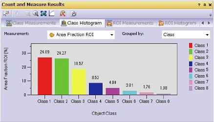
Grain classification results
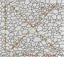
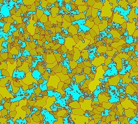
Grain Sizing in a Microstructure
Measure the grain size and analyze the microstructure of aluminum, steel crystal structures, such as ferrite and austenite, and other metals.
Supported standards: ISO, GOST, ASTM, DIN, JIS, GB/T
Microstructure of ferritic grains
|
| ||
Grain sizing intercept solution | Grain sizing planimetric solution with secondary phase |
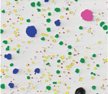
Evaluating Graphite Nodularity
Evaluate the graphite nodularity and content in cast iron samples (nodular and vermicular). Classify the form, distribution, and size of graphite nodes.
Supported standards: ISO, NF, ASTM, KS, JIS, GB/T
Ductile cast iron showing nodular graphite
Cast iron solution
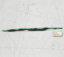
Rating Nonmetallic Inclusion Content in High-Purity Steel
Classify nonmetallic inclusions using an image of the worst field or worst inclusion found manually in the sample.
Supported standards: ISO, EN, ASTM, DIN, JIS, GB/T, UNI
Steel with nonmetallic inclusions
Inclusion worst field solution
Compare Images of Your Sample and Reference Images
Easily compare live or still images with auto-scaled reference images. This solution includes reference images in accordance with various standards (additional reference images can be purchased separately). Multiple modes are supported, including live overlay display and side-by-side comparison.
Supported standards: ISO, EN, ASTM, DIN, SEP
Steel with nonmetallic inclusions | Microstructure with ferritic grains | ||
|
|

Material Solution Specifications
| Solutions | Supported standards |
| Grain intercept | ISO 643: 2012, JIS G 0551: 2013, JIS G 0552: 1998, ASTM E112: 2013, DIN 50601: 1985, GOST 5639: 1982, GB/T 6394: 2002 |
| Grain planimetric | ISO 643: 2012, JIS G 0551: 2013, JIS G 0552: 1998, ASTM E112: 2013, DIN 50601: 1985, GOST 5639: 1982, GB/T 6394: 2002 |
| Cast iron | ISO 945-1: 2010, ISO 16112: 2017, JIS G 5502: 2001, JIS G 5505: 2013, ASTM A247: 16a, ASTM E2567: 16a, NF A04-197: 2004, GB/T 9441: 2009, KS D 4302: 2006 |
| Inclusion worst field | ISO 4967 (method A): 2013, JIS G 0555 (method A): 2003, ASTM E45 (method A): 2013, EN 10247 (methods P and M): 2007, DIN 50602 (method M): 1985, GB/T 10561 (method A): 2005, UNI 3244 (method M): 1980 |
| Chart comparison | ISO 643: 1983, ISO 643: 2012, ISO 945: 2008, ASTM E 112: 2004, EN 10247: 2007, DIN 50602: 1985, ISO 4505: 1978, SEP 1572: 1971, SEP 1520: 1998 |
| Coating thickness | EN 1071: 2002, VDI 3824: 2001 |
Sharing
Simple and Efficient Inspections
Use the GX53 microscope and PRECiV software to acquire images of diverse samples, conduct a variety of analyses, and generate professional reports.
> Click here for details about PRECiV
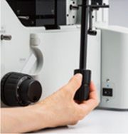
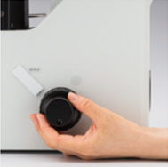
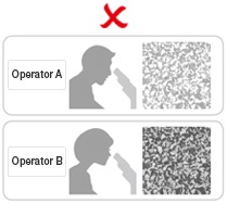
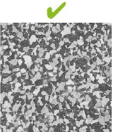
Easily Restore Microscope Settings: Coded Hardware
Coded functions integrate the microscope’s hardware settings with PRECiV image analysis software. The observation method, illumination intensity, and magnification can be recorded and stored with the associated images. The settings are easily reproduced so that different operators can conduct the same quality inspections with limited training.
|
|
|
|
| ||
Different operators use different settings. | Retrieve the device settings with PRECiV software. | All operators can use the same settings. | ||||


User Guidance Helps Simplify Advanced Analysis
The software guides users step-by-step through an inspection process that complies with the chosen industrial standard. Operators can conduct advanced analysis simply by following the on-screen guidance.
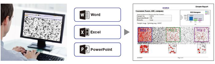
Efficient Report Generation
Creating a report can often take longer than capturing the image and taking the measurements. PRECiV software provides intuitive report creation to repeatedly produce sophisticated reports based on predefined templates.
Specifications
| Optical systemOptical system | UIS2 optical system (infinity-corrected) | |
|---|---|---|
| Microscope frame | Reflected light illumination |
Manual brightfield/darkfield selection by mirror unit
Manual field stop/aperture stop switch with centering Light source: White LED (with Light Intensity Manager) /12 V, 100 W halogen lamp/100 W mercury lamp/light guide source Observation mode: brightfield, darkfield, differential interface contrast (DIC)*1, simple polarizing*1, MIX observation (4 directional darkfield)*2 *1 Slider for exclusive use of this observation is required. *2 MIX observation configuration is required. |
| Microscope frame | Imprinting of scale | All ports reversed positions (up/down) from observation positions seen through the eyepiece |
| Microscope frame | Output front port (optional) | Camera and DP system (reversed image, special camera adapter for GX) |
| Microscope frame | Output side port (optional) | Camera, DP system (upright image) |
| Microscope frame | Electrical system |
Reflected light illumination
Built-in LED power supply for reflected light illumination Continuously-variable light intensity dial Input rating 5 V DC, 2.5 A (AC adapter 100–240 V, AC 0.4 A, 50 Hz/60 Hz) External interface (requires the optional BX3M-CBFM control box) Coded nosepiece connector × 1 MIX Slider (U-MIXR-2) connector × 1 Handset (BX3M-HS) connector × 1 Handset (U-HSEXP) connector × 1 RS-232C connector × 1, USB 2.0 connector × 1 |
| Microscope frame | Focus |
Rack and pinion with roller guide
Manual, coarse and fine coaxial handle; focus stroke 9 mm (2 mm above and 7 mm below the stage surface) Fine handle stroke per rotation: 100 μm (min. scale: 1 μm) Coarse handle stroke per rotation: 7 mm With torque adjustment ring for coarse focusing With upper limit stopper for coarse focusing |
| Tubes | Widefield (FN 22) | Inverted: binocular (U-BI90, U-BI90CT), tilting binocular (U-TBI90) |
| Nosepiece |
Brightfield Holes: 4 to 7 pcs, Type: Manual/Coded, Centering: Enabled/Disabled
Brighfield/darkfield Hole: 5 to 6 pcs, Type: Manual/Coded, Centering: Enabled/Disabled | |
| Stage |
Right handle stage for GX (X/Y stroke: 50 × 50 mm, max. load 5 kg)
Flexible right handle stage, left short handle stage (each X/Y stroke: 50 × 50 mm, max. load 1 kg) Gliding stage (max. load 1 kg) A set of teardrop and long hole types | |
| Weight | Approx. 25 kg (microscope frame 20 kg) | |
| Environment |
・Indoor use
・Ambient temperature: 5 to 40 °C (45 to 100 °F) ・Maximum relative humidity: 80% for temperatures up to 31 °C (88 °F) (without condensation) In case of over 31 °C (88 °F), the relative humidity is decreased linearly through 70% at 34 °C (93 °F), 60% at 37 °C (99 °F), and to 50% at 40 °C (104 °F). ・Pollution degree: 2 (in accordance with IEC60664-1) ・Installation/Overvoltage category: II (in accordance with IEC60664-1) ・Supply voltage fluctuation: ±10 % | |