Medical hypodermic needles
Measuring Up to Medical Hypodermic Needle Standards
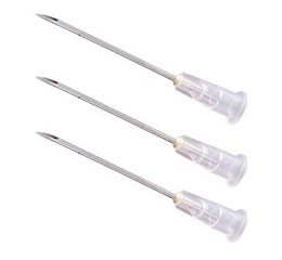
Hypodermic needles for medical syringes are inserted into the human body to inject drugs, collect blood or body fluids, and perform blood transfusions. Since the needles are inserted directly into the human body, they are strictly controlled under national and international standards that specify needle materials, shapes, and strengths. The standards specify hypodermic needle shapes for their lengths, outer diameters, and bevel angles. They also require that the needles have no flaws on outer and inner surfaces, contain no foreign matter, and have smooth finished surfaces.
Inspectors sometimes use multiple measuring instruments to meet these hypodermic needle standards, but extra equipment can make the workflow time-consuming and inefficient.
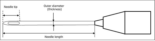
Diagram of a hypodermic needle
Benefits of Inspecting Hypodermic Needles Using the STM7 Measuring Microscope
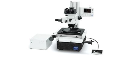
The Olympus STM7 measuring microscope enables inspectors to measure the dimensions and inspect the appearance of medical hypodermic needles without the need for extra measuring instruments. By using our measuring microscope with a CCD digital camera and STM7-BSW measurement support software, you can perform more efficient inspections.
Our STM7 measuring microscope helps you efficiently inspect hypodermic needles in three ways:
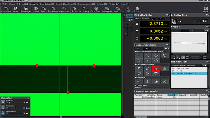
1. Measure the length of a hypodermic needle with flexible stages
Our measuring microscope measures length based on its stage’s travel distance. This means that, even when a hypodermic needle is longer than the field of view acquired with the objective, you can measure the needle length by moving the stage from one end of the needle to the other. The STM7 microscope is available with three stage travel distances, so you can measure the length of any medical hypodermic needle.
STM7-CS100 |
STM7-CS200 |
STM7-CS300 |
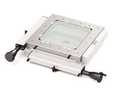
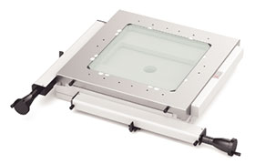
Three stages are available, each with a different measurement stroke
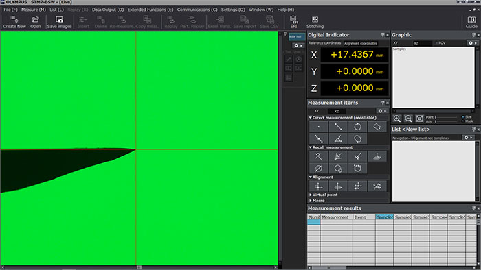
Needle length measurement
The stage moves straight in the X and Y directions. When taking measurements using the STM7-BSW software, you can set the desired coordinate axes. This way, you don’t have to put a hypodermic needle parallel to the stage to make accurate measurements.
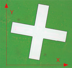 Original coordinate axes | 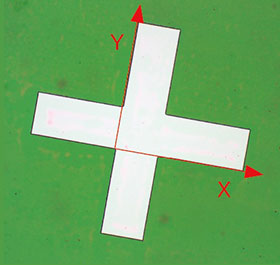 Newly set coordinate axes |
2. Measure hypodermic needle thickness and bevel angle with automatic edge detection
Needle thickness measurement
To easily obtain needle thickness data, click three points on the hypodermic needle edges, as shown above. (The actual user interface does not show the red points and lines.) To help you produce consistent measurement results obtained by different inspectors, the STM7-BSW software offers an automatic edge detection function. You can see how it works in the image below:
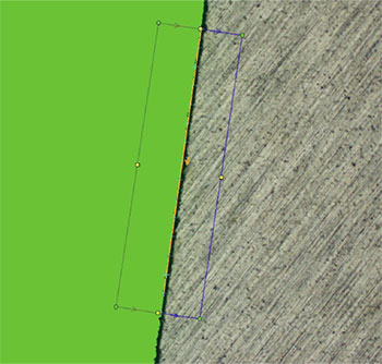
The software automatically recognizes any points in the box that have strong contrast between the sample and the illumination as an edge.
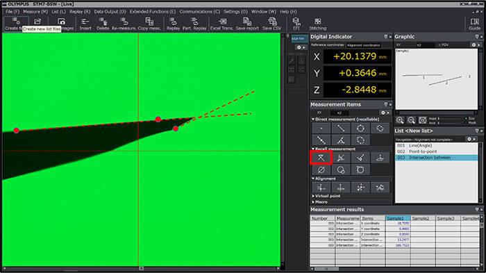
Needle bevel angle measurement
You can also obtain bevel angle data by clicking three points on the edges of the needle tip, as demonstrated in the image above.
3. Clearly observe the bevel of a hypodermic needle with extended focus
Reflected light illumination is used to inspect the appearance of a hypodermic needle’s bevel. But since the bevel is made by cutting a very thin tube diagonally, it is often difficult to bring it into focus. Our STM7-BSW measurement support software offers an optional extended focus function that brings the entire image into focus so that you can clearly observe the bevel.
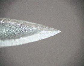 Only part of the bevel is in focus | 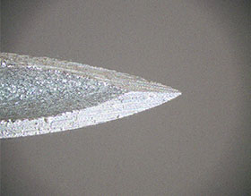 You can clearly observe the bevel with the extended focus function |