This summer, we announced the winners of our 2022 Global Image of the Year (IOTY) competition, an annual contest that recognizes the best in light microscopy imaging from around the world. For the 2022 edition, we went beyond life science images to include an exciting new category: materials science and engineering.
Now we’re happy to share more about the very first Image of the Year materials science award winner, Shyam Rathod from India. His winning image shows a unique subject—the crystal of a topical medicine for wart treatment. We spoke with Shyam to get a behind-the-scenes look at what it takes to capture a beautiful image like this so masterfully.
Q: What does your winning image show?
Shyam: The image shows the microscopic crystal of a topical medicine, a combination of salicylic acid and lactic acid, used to treat skin warts (also called a callus remover). It depicts the pattern that evolved during the crystallization process of the substance on the microscope slide.
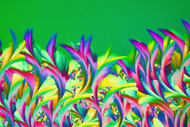
Winning image of the materials science and engineering category in Image of the Year 2022. Captured by Shyam Rathod from India. The image shows the crystal of a topical medicine for wart treatment named ABE, which is available in Poland. The dew was blown on a microscope slide with a straw and was captured in a single frame. A retarder and a two-cross polarization filter were used to bring out the colors.
Q: What do you find exciting about the image and subject matter?
Shyam: What excites me the most is that photomicrography provides a window to view the work of nature, as nature weaves different patterns in the space and time of the microworld. It is a blend of art and science, where a scientific medium is used to create an artwork.
The image obtained shows the beauty of the subject at the microscopic level, which mimics the real world in its beautiful patterns. Capturing an image like this not only requires scientific understanding and artistic acumen, but also the physical endurance and mental keenness to click the microscope camera at the exact moment of magic.
Q: How did you create the image?Shyam: Creating the image involved the preparation of slides and photography. Initially I put a drop of the callus remover, which is a combination of salicylic acid and lactic acid, on a slide. The drop was then pressed with another slide and gently moved to form a thin layer. As the layer dries, it starts to crystallize. I viewed the process of crystallization under the microscope. What was visible during the formation of the crystal was only a small part (around a 1 mm section) of the entire slide. I then imaged the regions of interest. The image was obtained using a camera mounted on the microscope by clicking at the very moment during the formation of the crystal when the beautiful pattern emerged. The microscope used was modified for polarization (two polarization filters, cut to size, were inserted in the optical path: one under the head and the other under the condenser). A microscope camera was mounted on the trinocular head. An Olympus LCPlanApo 20X objective was used as the objective. A DIY coupler was used, which was designed and fabricated on a 3D printer. |
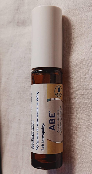
Callus remover used to create crystal artwork under the microscope. Image courtesy of Shyam Rathod. |
Q: What challenges did you encounter when capturing this image?
Shyam: The crystals of the medicine completely developed in a few seconds, so selecting the correct region to image during the tiny window of time was a challenge. Hundreds of test slides were prepared to obtain this image. Colors at different rotations of retarder gave different colorful results, and I chose the best one.
Another challenge was obtaining a microscope before starting photomicrography. I had to save carefully to procure a microscope. As there was no polarization in the microscope I bought, I had to apply a do-it-yourself approach with two polarization camera filters, cutting them to fit into the microscope. Low-budget cellophane plastic (retarder) was used to bring out the colors. The journey toward obtaining the final image has been strenuous, as the entire ecosystem was built over years.
Q: Why did you choose this image as your entry for the Image of the Year competition?
Shyam: This picture was obtained after immense effort. The texture, composition, and color pattern that emerged in the microworld, mimicking the outer world, was a magical moment captured with a camera. I chose this image as I found it to be the most beautiful one.
Q: What message about the image would you like to share?
Shyam: The fact that ‘nothing is constant in this universe, it’s continuously evolving’ can be observed during the crystallization process under microscope. Nature is so beautiful even at the microscopic level, one should have an eye to see it. Fortunately, we have microscopes and different lenses to peer into the microworld. I think microphotography is about harnessing the positive side of science, and it is one way in which science can be made popular among the masses and students. It could be developed as a hobby.
The microcrystal can be obtained using a chemical or the combination of various chemicals by dissolving the chemical in a solution or by melting the powder of the chemical on a temperature-controlled heater. There are a lot of possibilities. Different combinations of chemicals give different patterns, which keep changing in the space and time of the microworld.
Photomicrography of crystals is a rare art. I always feel like it’s an adventure going through the microscope into the microworld. It’s a rare and fulfilling experience, and a good way to spend both time and money. You must experience it for yourself.
Q: Where and when did you first learn to use a microscope?
Shyam: I was introduced to a microscope as a child at school. During my school days, I had purchased a small microscope to observe biological samples. Later, my use of the microscope was discontinued for decades only to resurface when I joined the Crystal Art Photomicrography group on Facebook. I also used to observe the celestial world with a telescope and take astrophotography images.
Q: When did you become inspired to use microscopes to create art? What first inspired you?
Shyam: I am thankful to the Crystal Art Photomicrography group and all the active members, especially Yogendra Joshi, for kickstarting my passion by sending me different chemicals to observe under the microscope. Looking at the microcrystal photographs of fellow members in the online group, I was inspired to continue my journey in the microworld. Most of my inspiration has come from the community, and the beauty of the artwork has kept my interest alive, turning it into my passion today.
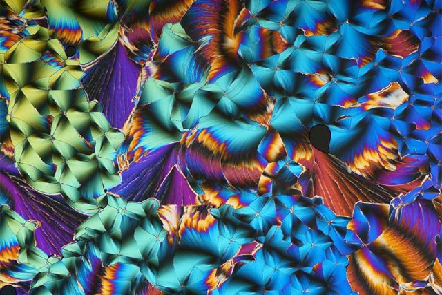
The crystallization of the callus remover captured under the microscope. Image courtesy of Shyam Rathod.
Q: How long have you been creating art with a microscope?
Shyam: I have been dedicated to pursing the art of crystal photomicrography for the past three to four years now. Prior to that, I had been mostly into observing biological samples with a microscope and celestial objects with the telescope.
Q: What do you find most fascinating about microscopy?
Shyam: Microscopy is a good way to utilize your time on this planet. It opens the window to the microworld. Everyone should have a chance to look at the microworld while busy in the bigger world. We have already spent a lot of resources to create elements with the potential to destroy. Microscopy is an affordable solution to observe the process of creation. I believe it can regenerate good in the world, as different minds may discover different findings that could enrich the knowledge of humanity. That is what makes microscopy fascinating, apart from the beauty and thrill involved in it.
Q: What do you do professionally, and what is your educational background?
Shyam: I work for the Maharashtra State Electricity Transmission Company (MSETCL) as the deputy executive engineer. MSETCL is a wholly owned state transmission utility (STU) under the Maharashtra Government that is responsible for the transmission of electricity. I hold a bachelor’s degree in electrical engineering from Veermata Jijabai Technological Institute (VJTI), Mumbai University (India).
Q: Does your profession intersect with imaging, or is photomicrography more of a hobby, art form, or passion for you?
Shyam: My profession doesn’t intersect with imaging. I have been doing photomicrography as a passion after work and on weekends on a continuous basis. My profession is my duty, and microphotography is my passion as I seek happiness from it.
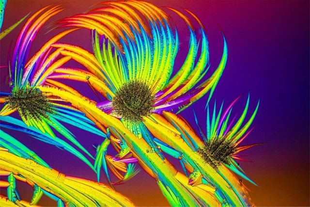
Microscope image showing the melting of paracetamol and urea captured under polarized light with a camera flash. Image courtesy of Shyam Rathod.
Q: What are you are currently working on professionally and artistically?
Shyam: In my photomicrography, I have been working with different chemicals for making crystals. I am turning slowly toward making videos of crystal growth using different techniques and chemicals.
Q: Is there anything else you’d like to share about your experience in the Image of the Year contest?
Shyam: I am very excited to receive the SZ61 stereo microscope as the prize for the Image of the Year materials science award. I would have never been able to get a hold of such a fine microscope. It is precise and great for imaging.
I highly appreciate the work being done by Evident, as the art hidden in the microworld obtained in the small setting of a house in a small town could be made known to the world! This Image of the Year contest will stand as an inspiration to many. It may also provide a solution to those who, like me, might be struggling to get a hold of the right microscopy device to make their dream come true.
Art of Materials Science—Be Part of the Next Image of the Year Contest
Our next Image of the Year contest is just around the corner! If you’re interested in creating and sharing your own materials science and engineering images, join our e-newsletter to be first to hear details on our next imaging competition. To get inspired, head to our Image of the Year page to download Shyam’s winning image as a stunning wallpaper for your desktop and
smartphone.
Related Content
3 Microscopy Techniques that Transform the Ordinary into the Remarkable
The Potential of AI-Based Image Analysis in Metallography and Materialography
Discovering the Unseen Beauty of Sand Under a Digital Microscope