1. Principles of Laser Scanning Microscopes
Introduction
One of the factors that contributes to the recent considerable reduction in size and high integration of electronics and other devices is miniaturization of the electronic components that make them up. In addition, new fine functional materials have been developed and widely used in industrial fields including the automobile, aviation, metal, and chemical industries. As a result, higher accuracy and resolving power are required for minute three-dimensional measurement of these components and materials. Although a variety of devices are available to satisfy these requirements, the confocal microscope is gathering attention as the device of choice for easy three-dimensional surface profile measuring in air, without touching the sample. Covered within are the basic principles of and the features and applications of Olympus industrial laser scanning microscopes, with primary focus on the reflection-type confocal laser scanning microscope (hereafter referred to as the laser scanning microscope), which is aimed at high resolution detection
1-1. Confocal Optical System
Of the principles of laser scanning microscopes, the most basic is the confocal optical system. The image formation optical system in a typical optical microscope and the confocal optical system are shown in Figure 1. The confocal optical system has a pinhole with a circular opening at the position that conjugates the focal position of the objective lens (image forming position) to only detect light at the focused position. Although it is important to evenly illuminate a certain area for a typical optical microscope, in the confocal optical system the light emitted from the point light source is radiated by the objective lens so that the light converges to one point on the sample. In general, a specific wavelength of laser light, which travels in a very straight line, is used as the point light source so that a large amount of light converges at one point. This reduces unnecessary scattered light from the environment and improves contrast compared with ordinary optical microscopes, which evenly illuminate the sample. The light reflects on the surface of the sample, goes back along the same optical path, is separated by the beam splitter, and converges on the pinhole. This optical system is able to obtain information from only the focal position as most of the reflected light from places other than the focal position is blocked at the pinhole. This means that the confocal optical system can act like a height sensor because of its narrow depth of focus while a conventional optical system cannot due it's large depth of field which includes unfocused data. In addition, while out of focus images from places other than the focal position are superimposed in an ordinary optical microscope, the confocal optical system blocks the reflected light from places other than the focal position at the pinhole, forming a perfectly focused clear image with good contrast. Although it is said that the diameter of the pinhole in the confocal optical system should be smaller than the divergence caused by the diffraction of the beam spot, this diameter is determined by balancing the sensitivity of the detector because the amount of light is reduced as the pinhole diameter is narrowed.
Figure 1: Image Formation Optical System and Confocal Optical System for Optical Microscopes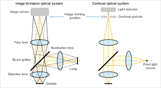
1-2. Two-Dimensional Scanning
As described above, the confocal optical system only obtains information in the direction of the optical axis. Therefore, some kind of two-dimensional scanner system orthogonal to the optical axis is required to convert data into images. Since this two-dimensional scanner system uses raster scanning to create an image, the accuracy of this scanning system directly determines imaging performance; accurate two-dimensional scanning is one of the most important technologies used in laser scanning microscopes. Scanning systems are generally divided into the sample scan method and the laser scan method. The sample scan method is performed by scanning the XY stage on which the sample is placed. This method allows the user to create an image of a large area without the need for optical scanning function, therefore, the confocal optical system will have a simpler structure. In addition, the XY stage can be accurately driven relatively easily. However, it takes a very long time to capture data and at a high resolving power it is necessary to drive the XY stage, including the sample for observation, with great precision. The laser scan method is performed by scanning a laser beam onto a sample in two dimensions, X and Y, using two scanning mechanisms. While the sample scan method is appropriate for capturing large waves on the sample surface, the laser scan method is ideal for capturing minute shapes. Most industrial laser scanning microscopes generally employ the laser scan method to put priority on image acquisition time. For example, an acousto-optic deflector (AOD), polygon mirror, or resonant galvano mirror is used in the X direction as the laser scanning mechanism because high-speed scanning is required. These scanning mechanisms are briefly introduced below.
1-2-1. Acousto-Optic Deflectors (AOD)
An acousto-optic deflector (AOD) is an element that makes use of the diffraction of light. When modulating the frequency of ultrasonic waves and applying it to a material, the change of refractivity in the material functions as the diffraction grid, providing an appropriate light deflection angle. Although an AOD can scan very quickly, the scanning range is limited. In addition, a configuration that leads to descanning (re-incidence of reflected light from the sample) must be avoided as descanning greatly decreases efficiency. For this purpose, adding a line sensor or other technique is required. It is also important to exercise sufficient care when using an AOD because the lens effect can cause astigmatism.
1-2-2. Polygon Mirrors
Polygon mirrors have a very simple configuration and are used in many fields that require laser scanning. The laser is scanned by putting a mirror on each face of a polytope and rotating the polytope at high speed using a motor or other device. The scan speed and angle of the laser is determined based on the number of mirror faces (assuming that the rotation speed is constant). Extremely precise adjustment is required because the rotation accuracy determines the scan accuracy.
1-2-3. Resonant Galvano Mirrors
A resonant galvano mirror is a compact laser scanning mechanism that supports relatively large oscillation angles. Because its speed is determined by mechanical resonant frequency, this mechanism is limited in terms of speed compared to other scanning mechanisms. However, recent resonant galvano mirrors can acquire several one-megapixel images every second. Mirrors manufactured with MEMS (micro-electro-mechanical systems) technology have also been developed, allowing for a reduction in device size. MEMS scanners are a combination of movable plate, torsion (I think you need a glossary)bar, and support frame made by etching a single monocrystalline silicon board. The movable plate has coils driven by a magnetic circuit. Two-dimensional scanning can be accomplished by taking advantage of the characteristics of a high-speed scanning mechanism and combining it with a relatively low-speed scanning mechanism in the Y direction. A nonresonant galvano mirror is often used in the Y-direction scanning mechanism partly as a matter of convenience.
1-3. Confocal Effect and Extended Focus Image
Figure 2: I-Z Curve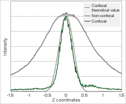 | Figure 2 illustrates the effect of the above-mentioned confocal optical system. The vertical axis of this figure shows the output from the detector after the light passes through the pinhole. The output was acquired while moving the sample and objective lens relative to each other in the Z direction, without two-dimensionally scanning with the confocal optical system, in order to confirm this effect. The horizontal axis indicates the travel distance in the Z direction. This waveform is generally called an I-Z curve. Comparing the non-confocal output with the pinhole removed and the confocal output acquired under the same conditions reveals that the confocal optical system shows a steep waveform. An image of a stepped sample where all height positions are in focus (extended focus image) can be obtained by scanning the laser light in two dimensions, relatively moving the sample and objective lens, and saving the brightest light intensity value of each pixel on the image to make use of this characteristic. Figure 3 shows how the sample image is actually captured. When scanning the laser light across the top face in the figure in two dimensions and focusing it, blurry images are eliminated due to the characteristics of a confocal optical system. Therefore, the square part is captured. When the laser light is moved in the Z direction and the second face is focused on, the smallest L-shaped part is created. By sequentially repeating this step and then capturing and overlapping the image of each face, an image with a deep focal depth in which every face of the sample is brought into focus at high resolving power in the horizontal direction (extended focus image) can be created. |
Figure 3: Extended Focus Image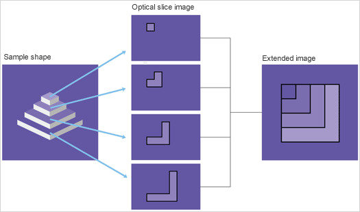

1-4.Three-Dimensional Images
In the confocal optical system, the Z position with the maximum intensity, that is, the brightest Z position indicates the height information of the sample surface at that point. Taking advantage of this fact, the height information of the sample can be captured by recording the Z position with maximum intensity. Figure 4 shows how height information is actually captured. In the same way as when capturing an extended focus image, move the sample and objective lens relative to each other and
save the information on the brightest Z position for each pixel, where the maximum intensity can be obtained, while moving from height Z1 to Z2. This makes it possible to obtain the surface profile of the sample in the image acquisition area. A variety of analyses can be performed based on this information. This means that both intensity and height information can be obtained at the same time by capturing the maximum intensity value of each pixel into the extended image memory and the height at
that time into the height image memory. This is the most significant characteristic of confocal microscopes that sets them apart from other microscopes.
Figure 4: Capture of the Height Information of an Image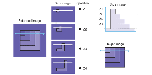
- 1. Basic Principles of Laser Scanning Microscopes
- 2. Resolving Power of Laser Scanning Microscopes
- 3. Measurement Accuracy of Laser Scanning Microscopes
- 4. Software for Laser Scanning Microscopes
- 5. Basic Features of Olympus Industrial Laser Scanning Microscopes
- 6. Application Examples
> Click here for detail about OLS5000, laser confocal microscopes
> Click here for detail about OLS4500, nano search microscopes