Annegret Janovsky is a senior specialist for industrial microscopy at Olympus who uses her imaging expertise for a unique hobby: microscopic sand photography. In her spare time, she collects sands from beaches around the world to produce beautiful, detailed images of their grains using our DSX1000 digital microscope.
DSX1000 digital microscopes are typically used for failure analysis in the manufacturing industry and can identify corrosion or fractures in materials. Its key advantages for these analyses include:
- Superior resolution at high magnification
- Long working distance objectives
- The ability to use multiple observation methods at all magnifications
For Annegret, the same advantages that the DSX1000 digital microscope brings to failure analysis make these systems ideal for imaging sand. The result is stunning images that capture the variation in grains from beaches worldwide.
We caught up with Annegret to ask her about some of her favorite images.
1. Pure mineral sand from the Scottish Highlands
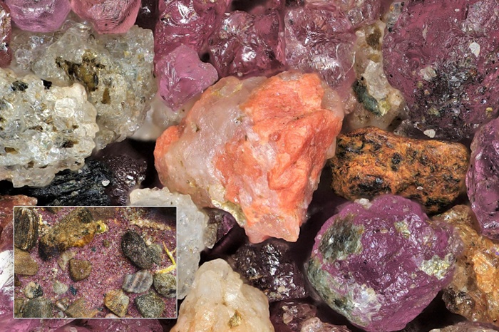
The first image shows a pure mineral sand from Loch Achilty in the Scottish Highlands.
Captured under the microscope in 2015, this sand is particularly special to Annegret because it is the first one that she imaged.
She explained how she discovered the sand.
“I was looking for good samples to test the image quality of a new range of microscopes, when I remembered a beautiful violet sand that I encountered while travelling in Scotland with my husband over 20 years ago,” Annegret commented.
She continued, “This prompted a long journey back to Scotland to find it! After two days of searching, we found the sand on the banks of the loch and collected a sample. The resulting images taken with the DSX were exceptional and motivated me to start collecting sand from around the globe.”
2. Organic sand from Taketomi Island
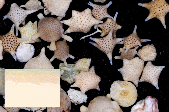
The second image shows an organic sand from Taketomi Island—a Japanese island in the East China Sea. It is composed of the shells of forams—microscopic organisms that form hard shells of various shapes.
“These grains are particularly amazing as the whole shell is formed by a single-celled organism,” Annegret explained. “The shells are relatively large at 2–3 mm each, so this image was taken using a 3X XLOB objective from Olympus.”
Annegret discussed why these objectives are so beneficial for her microscope artwork.
She said, “These objectives are useful for 3D samples because they combine the resolution of standard material objectives with very long working distances—reducing the risk of sand grains in the optics!”
3. Heavy mineral sand from Canada
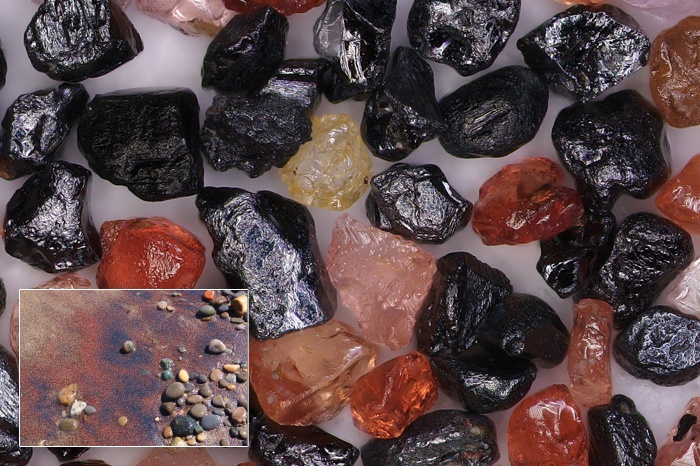
The third image shows a heavy mineral sand from Lake Ontario, Canada. It is composed mainly of garnet grains (red) and magnetite grains (black).
Annegret explained her technique for capturing these colorful grains in detail.
She said, “This sand was imaged using a 10X XLOB objective. As with the first image, these grains are small and have interesting colors. From my experience of imaging different materials, I knew that darkfield microscopy is best for imaging natural colors. It is also useful to have a light source above the sample to capture surface details. The DSX1000 allows simultaneous brightfield and darkfield illumination from different directions, which is ideal for these types of samples. In fact, all of the images featured here were taken using MIX illumination.”
4. Organic sand from Ireland
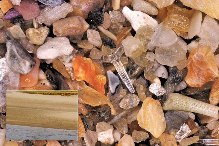
The final image shows an organic sand with a heterogenous composition collected from Lahinch in Ireland.
Annegret explained what makes this sand so unique.
She said, “This type of sand is different from the others in that it is a heterogenous mix of purely biogenic components. It is impossible to find the sources of all these grains—they could be shells, forams, or other organic matter. This particular sample contains part of a sponge as well as a broken sea urchin spine.”
“A crucial advantage to using the DSX1000 for imaging sand is its ability to acquire Z-stacks—all of the images here were acquired using focus stacking,” Annegret explained.
A Growing Sand Collection from Coastlines Around the World
Annegret now has over 300 different sands in her collection, and the number keeps growing.
“A lot of family and friends have heard about my hobby and are sending me sand from around the world,” Annegret said. “I have some exciting plans to image sand from hydrothermal vents and volcanic areas in the near future.”
She added, “Sand is surprisingly beautiful, and I think sand will be a passion of mine for my whole life!”
To learn more about the DSX1000 system’s various applications and its advantages over conventional digital microscopes, check out our digital microscopy resource page.
Related Content
Imaging on the Fly—Observing Medically Important Insects Using Digital Microscopes