![]()
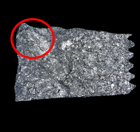
A fractured metal surface
Fractography
Fractography is an analytical technique used to determine the cause of fracturing in a metal structure. The method relies on the analysis of dimples and slips on the fracture’s surface to determine whether it was caused by corrosion fatigue, stress corrosion, or a stress fracture caused by fatigue, brittleness, ductility, or creep. The direction of the break can also usually be determined, providing valuable information about the amount of stress and load that the metal part can withstand.
Fractography has become increasingly important as infrastructure continues to age and quality control issues cause problems. Optical or digital microscopes are essential fractography tools that are used to capture high-quality images for analysis. However, fractured surfaces can have complex shapes, making their inspection using a microscope challenging.
4 Challenges of Inspecting Fractured Metal Surfaces with a Microscope
Observing a Fracture Surface Over a Wide Area
When starting an analysis, inspectors observe the surface with the widest field of view possible to find the cause of the damage. One solution is to use stitching where multiple images are combined to form one large image. However, poor quality optics can cause the seams between the images to be visible, making it challenging to locate the cause of the damage.

Seams between overlapped images are highly visible
Keeping Track of the Observation Position During Analysis
The features and topography of a fractured surface can look very similar, making it easy to lose your position during the analysis. If a user loses their position, they may have to start over.
Difficulty Locating a Singularity on the Fracture’s Surface
Low-magnification observations are conducted to locate singularities. When there are height differences on the surface, the microscope’s depth of focus can make it impossible to completely focus the image, even under low magnification. When this happens, inspectors can use an image processing technique called focus stacking to create a clear image. However, this process can be slow, and if a different section of the sample needs to be examined, the user must return to the live image to navigate.
The ability to locate the singularity also depends on the resolution of the microscope’s objective lens. Most low-magnification lenses have a lower resolution, which can make it difficult to capture an image of a singularity on a complex-shaped fracture surface.
Difficulty Analyzing a Singularity
Once a singularity has been located, it is examined under high magnification. As discussed above, the objective’s resolution significantly impacts image quality, so if an inspector attempts to zoom in on the singularity, the image can be blurry. When this happens, the inspector needs to change to a higher magnification lens with a better resolution. However, this process means that the image needs to be refocused and the singularity reacquired, which adds time to the inspection.
Another issue with high-magnification objective lenses is that they have a shallower depth of focus, making the focus-stacking technique to obtain a fully focused image problematic. It is also possible that, due to the small working distance, the lens can collide with the sample when attempting focus stacking, potentially causing damage to both.
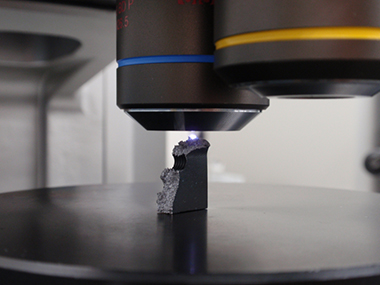
Shallow depth of focus
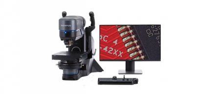
Advantages of the DSX1000 Digital Microscope for Fractography
DSX low-magnification objectives combine a deep depth of focus with high resolution to provide sharp images. And if you need an even deeper depth of focus, press the Focus Depth Up button on the console. These features enable you to display clear images and find singularities more quickly. If the fracture surface has large irregularities, you can still obtain high-resolution images.
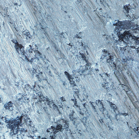
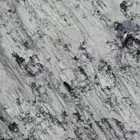
In the image captured using the DSX1000 digital microscope (right), the topography of the fractured piece is much clearer than the image captured using a conventional microscope.
Conventional: 31X magnification |
DSX1000: 30X magnification |

High Magnification
Switching to high magnification for detailed inspection on the DSX1000 digital microscope is simple. The quick-change objectives enable you to slide out the low-magnification lens and replace it with a high-magnification one without losing your position on the sample.
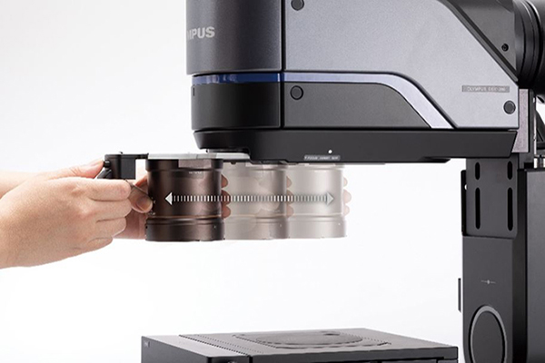
The objectives are mounted to quick-change cartridges that slide in and out of the zoom head.
Like the low-magnification lenses, the high-magnification DSX objectives also combine high-resolution and a deep focal depth so that, during a detailed inspection, you can view the fracture surface with in-focus images. The Focus Depth Up function also works with the high-magnification lenses when a deeper focal depth is required.
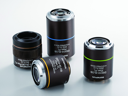
XLOB series objectives
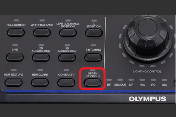
Depth of Focus Up button
To correct for the issues commonly found during image stitching, such as visible borders between the images, the DSX1000 digital microscope offers an improved algorithm for pattern matching and shading compensation. The result is higher quality stitched images without position shifts or uneven image seams.

The system’s macro map feature shows the observation position to help users better understand where they are looking on their sample. Now, users can continue their observation without losing sight of their position.
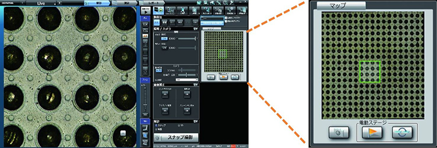
The macro map shows you the observation region on your sample.
Comparing Images Captured with the DSX1000 and a Conventional Microscope
In the image captured by the DSX1000 digital microscope, you can observe the characteristic state of the fracture surface at a high resolution.
*To guarantee XY accuracy, calibration work must be undertaken by an Olympus service technician.
|
|