BXiS
Discontinued Products
Overview
This product has been discontinued, check out our current upright microscopes >>
Olympus BXiS microscopes were developed based on cutting edge optical and imaging technologies to address various materials science applications. The combination of BXiS and OLYMPUS Stream software streamlines workflows and offers flexible solutions for image acquisition, measurement and reporting.
- Olympus Is Dedicated to Making Microscope System Solutions to Support Your Work on All Levels
- OLYMPUS Stream Software Adapts to Your Every Requirement
- The BXiS Simplifies Your Image-Acquisition Workflow
- BXiS Offers the Perfect System for Your Sample and Solution
- Olympus Broad Range of Digital Cameras
- From Simple Measurements to Complex Image Analysis
- BXiS Expands to Meet Your Future Needs
Olympus Is Dedicated to Making Microscope System Solutions to Support Your Work on All Levels
Keep Your Workflow Streamlined
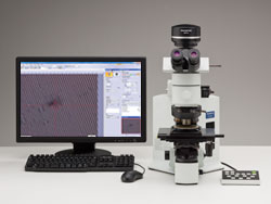
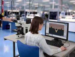 BX51M and OLYMPUS Stream | Your time is just as important as your working conditions. That's why the BXiS system's imaging and control software can be personalized to fit your process flow. An easy-to-use interface guides you effortlessly through every step from image adjustment image capture, measurements, report creations and data basing or whatever you need to achieve. As a result, you'll find that you can complete your tasks more efficiently regardless of their complexity. |
Build Your Olympus System Your Way
The fully adaptable BXiS concept shows how we're able to offer manual or automated systems that fit your needs and budget. Create and tailor your system however you wish!
Easily Expandable To Future Applications
With the BXiS system, you're prepared not only for today's applications but for tomorrow's as well. Whatever the future may bring, you'll be ready to keep up with advances in technology with Olympus BXiS system solutions.
OLYMPUS Stream Software Adapts to Your Every Requirement
User Interface Function
 OLYMPUS Stream user interface | As you progress from image capture to report creation, the tool windows you need are always displayed at every stage. You can quickly and easily access control parameters. |
 Work flow 1 : Image Acquisition | 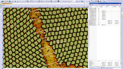 Work flow 2 : Measurements |
 Work flow 3:Objective Detection | 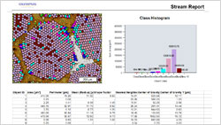 Work flow 4: Report Generation |
Dynamic User Interface
Depending on your needs, you can arrange the layout of the tool window to best fit your workflow. You can create customized layouts with all the necessary functions for each activity.
Microsoft Office Integration
OLYMPUS Stream offers complete integration with Microsoft Word and Microsoft PowerPoint.* This allows for the creation of professional reports and presentations accommodating your company templates including your own headers and footers. OLYMPUS Stream uses a dedicated menu and toolbar that is integrated into your preferred Microsoft Office package for an improved image handling experience. Tables and graphs can also be easily exported into Microsoft Excel for extended processing. Due to a unique compression method, OLYMPUS Stream ensures your report files are a reasonable size for easier data exchange by email.
*Microsoft PowerPoint Assistant is optional
My Function
You can create intuitive workflows based on the most frequently used functions, simplifying repetitive tasks so that even new users are able to operate the software easily and efficiently.
The BXiS Simplifies Your Image-Acquisition Workflow
Magnification Readout
Accurate measurements depend on correct magnification settings. The BXiS provides this critical capability automatically with a manual coded system or via a fully motorized configuration. The objective lens settings are automatically reflected in the scale and measurement results displayed on the monitor and output to your reports.
 Coded or motorized nosepiece ensure correct magnification calibration. |
Quality Control and Traceability
For quality control and traceability purposes, the OLYMPUS Stream software creates a calibration report for important information, such as magnification and calibrated pixel values, after the initial installation. The Info Stamp created from updated calibration information can be overlaid on the acquired image and report creation.
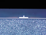
Instant Extended Focus Image (EFI)
When combined with the OLYMPUS Stream software,Instant Extended Focus Image provides easy images for samples that extend beyond the depth of focus. The manual EFI lets you use the smooth focus adjustment to combine many images in the z axis, providing you with one combined output that can be used for visualization or measuring in x and y.
 Normal image | 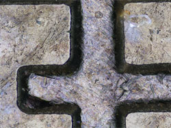 EFI image |
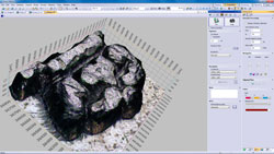
Manual Multiple Image Alignment (MIA)
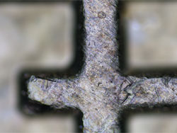
 Multiple Image Acquisition of Platinum Wires Cross Sections | OLYMPUS Stream software provides Manual Multiple Image Alignment to enable the creation of panoramic images of samples that extend beyond the field of view. The simple step-by-step process quickly allows you to combine the images. The OLYMPUS Stream software then rapidly stitches them together, providing you an output ready for simple visualization or complex measurement. |
BXiS Offers the Perfect System for Your Sample and Solution
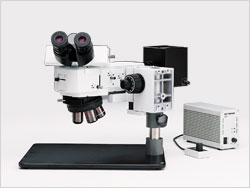
BXiS Motorized Microscopes - BX61
 BX61TRF/BX-RLAA/U-AFA2M/DP Series | With the BX61 microscope, complex operation settings such as illumination level, lens selection and aperture setting, can be set to operate from a single key, either on the microscope keypad or via the PC. This feature makes it easy to reproduce observation conditions with the touch of a single button. A variety of motorized modules, including nosepieces and illuminators, are available to provide you with the full flexibility of the BXiS. |
BXiS Microelectronics Inspection Microscope - BX41M-LED
 BX41M-LED/BX-AKMA-LED/DP22 | The BX41M-LED has an ESD dissipation capability that protects electronic devices from static electricity, the human body, or nearby environments found in the laboratory or shop floor. |
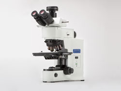
BXiS Modular Microscope - BXFM
 BXFM/BXFM-ILH/BX-RLA2 | The BXiS can also be adapted to special applications or integrated into other instruments. The modular construction provides for straightforward adaptation to unique environments and configurations with a variety of special small illuminators and fixturing mounts. |
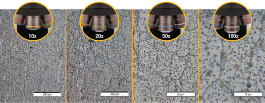
Choose from a Range of Objective Lenses to Customize Your System
Olympus offers a wide variety of objective lenses to suit every observation technique. You can select the right lens for your application from our family of over 150 objective lenses. Color fidelity is important for accurate, efficient inspection. The UIS2 objective lens series yields natural color reproduction by combining carefully selected high-transmittance glass and advanced coating technology.
| Example Observation Images | |||
 Magnetic Head (Brightfield) | 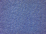 DVD (Brightfield) | 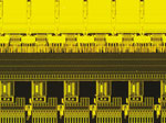 Wafer (Fluorescence) |  Color Filter (Transmitted) |
| Diverse Lineup Allows Selections According to the Purpose | |
| MPLAPON | Plan Apochromat Objective Lens Series |
| MPLFLN | Semi Apochromat Objective Lens Series for Brightfield |
| MPLFLN-BD | Semi Apochromat Objective Lens Series for Brightfield and Darkfield |
| MPLFLN-BDP | Semi Apochromat Objective Lens Series for Brightfield and Darkfield |
| LMPLFLN | Long WD Semi Apochromat Objective Lens Series for Brightfield |
| LMPLFLN-BD | Long WD Semi Apochromat Objective Lens Series for Brightfield and Darkfield |
| MPLN | Plan Achromat Objective Lens Series for Brightfield |
| MPLN-BD | Plan Achromat Objective Lens Series for both Brightfield and Darkfield |
| SLMPLN | Super Long WD Plan Achromat Objective Lens Series |
| LCPLFLN-LCD | Long WD Semi Apocromatic Objective Lens Series for LCD |
| LMPLN-IR/LCPLN-IR | Long WD Plan Achomatic Objective Lens Series for Near Infrared light |
> Click here for details about UIS2 objective lenses
Olympus Broad Range of Digital Cameras
DP73
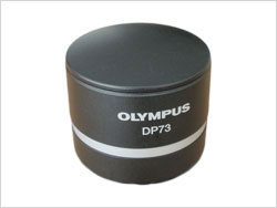 | The DP73 is a 17.3 megapixel cooled digital color camera for microscopes with pixel-shift echnology to attain ultra high resolution, high sensitivity and wide dynamic extended range function to produce contrast balanced images. |
DP27
The DP27 is a 5 megapixel digital camera for microscopy with high-fidelity color reproduction and progressive scanning free from color shift, providing live images with superb quality, seamlessly integrated into the OLYMPUS Stream analysis software
DP22
This 2.8M-pixel color CCD camera can be controlled from a space-saving, intuitively operated control box that incorporates the 12 most frequently used measurement functions for efficient inspection of industrial parts.
> Click here for the Olympus' lineup of digital cameras.
> Click here for the comparison sheet of Olympus' digital cameras.
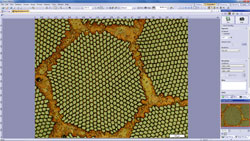
From Simple Measurements to Complex Image Analysis
Measurement
For quality control and inspection, measurement is an essential function. Even the entry-level OLYMPUS Stream Start includes interactive measurement functions such as distances, angles, rectangles, circles, ellipses, and polygons. All measured results are saved with the image fi les for further documentation.
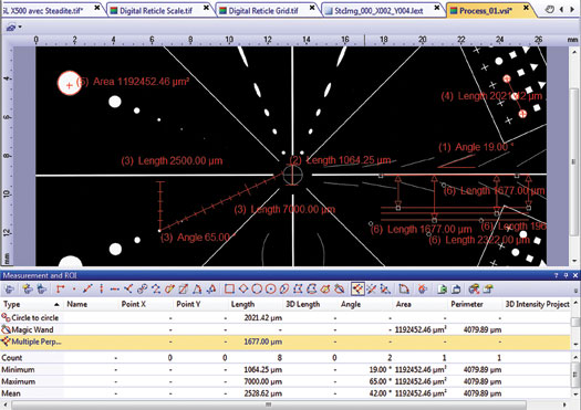
Measurement options
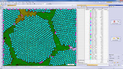
Count and Measure
Object detection and size distribution measurement are among the most important applications in digital imaging. OLYMPUS Stream incorporates a detection engine that utilizes threshold methods to reliably separate objects (e.g., particles, scratches) from the background.
Materials Science Solutions
OLYMPUS Stream offers an intuitive, workflow-oriented interface for complex image analysis. At the click of a button, the most complex image analysis tasks can be executed quickly, precisely, and in compliance with most common industrial standards. With a significant reduction in processing time for repeated tasks, materials scientists can concentrate on analysis and research. Modular add in for inclusions and intercept charts are easily added at any time.
BXiS Expands to Meet Your Future Needs
Database Management
When you need to efficiently browse through thousands of images and other files created in the past, OLYMPUS Stream can streamline your workflow from image capture through data management.
The software incorporates a client-server database based on Microsoft SQL Server Express. It allows you to assign user-definable fields (creation date, project ID, parts number, deadline, and metadata) into image and other files and folders, permitting efficient data sharing and quick searches. With the flexible right-setting capability, you can confidently handle the data.
Simple Network Connections
The OLYMPUS Stream Netcam solution lets any authorized network user connect to your OLYMPUS Stream PC and visualize the same image in real time with a web browser. What’s more, the DP22 also provides self-contained integration into your local network, allowing you to share your work across your office or around the world.
Microsoft Office 2013
Stay up to date with OLYMPUS Stream. The report tool and presentation assistant can use the latest version of Microsoft Office suite.
* Microsoft Office 2007, 2010 are also supported.
Windows 8.1
Microsoft Windows 8.1 (32bit/64bit) is the new standard in operating systems. OLYMPUS Stream fully utilizes all of the advanced capabilities for your everyday tasks.
* Microsoft Windows 7,8 is also supported.