Kompaktes Polarisierungsmikroskop CX31-P
Abgekündigte Produkte
Überblick
This product has been discontinued, check out our current upright microscopes >>
The CX31-P microscope is ideal for polarized light observations. Compact in design , yet capable of technically advanced tasks such as retardation measurements , the CX31P is a wise choice for daily laboratory use.
High Optical Performance
Space Saving High Performance Polarizing Microscope
CX31-P space saving body offers high quality polarized images and a wide range of options.This model is a cost-effective solution with a versatility of functions.
| Example Observation Images | |||
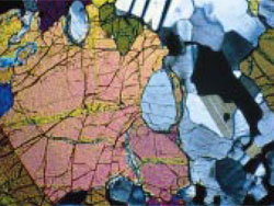
 Biotite Granite | 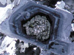 Quartz Diorite |  Tencel |  Sodium Urate |
Objective Lenses Dedicated to Polarized Light
Choose from specially manufactured strain-free objective lenses (PLN4xP, ACHN-P and UPLFLN-P) that are designed to ensure the highest quality images during polarized observations.
 UPLFLN-P Series |
| |||||||||||||||||||||
 ACHN-P Series |
| ||||||||||||||||||
Improved Polarizer and Analyzer
Specially designed condenser and polarizing filters offer, high EF value* and provide greatly enhanced polarized light images.
* EF value: the ratio of brightness of parallel Nicols to crossed Nicols. The higher is the EF value, the less is the distortion of the optical system. This means that a higher EF value is superior in polarization characteristics.
Orthoscopic/ Conoscopic Observation
Various operations are centralized including switching between the conoscopic and orthoscopic methods by the insertion/removal of a Bertrand lens.
 Peridotite (crossed polarizers) | 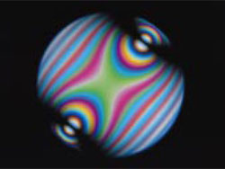 Conoscopic image of a biaxial crystal (topaz) |
High-rigid Frame that Prevents Image Blurring
To make the most of the superior optical performance, the basic performance has been enhanced with a highly rigid frame, interior focus mechanism and stage.
Binocular Observation Tube that Allows Interpupillary Adjustment without Precise Reticle Alignment
The binocular observation tube has a mechanism that allows interpupillary adjustment without inclination of the reticle such as a cross-hair in an objective lens. This enables exact alignment of the vibration direction of polarization.
Accessories Supporting High-level Polarized Observation
Precise Centering
The circular rotatable stage has two centering knobs and allows smooth sample rotation. A selectable click stop every 45 degrees enables accurate observation and measurement.
Picture of Rotating Stage
 X-Y Mechanical Stage | For added versatility an x-y mechanical specimen holder is available for easy x-y movement of one's specimen. Mounting an attachable cross movement mechanical stage (U-FMP) onto the circular rotatable stage allows precision x-y focus, especially useful at higher magnifications. Interference between the mechanical stage and the objectives is eliminated, and images of superb quality can be effortlessly observed with all objective magnification. |
 Objective Lens Centering Adapters | Four objective centering adapters center your objective for precise specimen observation. |
Wide Choice of Compensators
| Retardation Measuring Ranges of Various Compensators | ||
| Compensator | Measuring Range | Major Application |
| U-CTB Thick Berek | 0-11,000nm | Measurement of Large Retardation (R*>3λ), (Crystal, Giant Molecule, Fiber, Photoelastic Distortion, etc.) |
| U-CBE Berek | 0-1,640nm | Retardation Measurement (Crystal, Giant Molecule, Fiber, Body Tissue, etc.) |
| U-CSE Senarmont | 0-546nm |
Retardation Measurement (Crystal, Body Tissue, etc.)
Contrast Intensification (Body Tissue, etc.) |
| U-CBR1 Brace-Kohler 1/10λ | 0-55nm |
Minute Retardation Measurement (Crystal, Body Tissue, etc.)
Contrast Intensification (Bbody Tissue, etc.) |
| U-CBR2 Brace-Kohler 1/30λ | 0-20nm | |
| U-CWE2 Quarts Wedge | 500-2,200nm | Preliminary Measurement of Retardation (Crystal, Giant Molecule, etc.) |
The concurrent use of interference filter 45IF546 is recommended for improving measuring accuracy. (Except U-CWE2)

Test Plates/Compensators
Easy Adaptation of Digital Cameras, Image Analysis Software and Automated Accessories
The OLYMPUS Stream Image Analysis Software has been designed specifically for industrial microscopy applications with intuitive menus and advanced software routines. Users are empowered with the latest image analysis and management solutions to satisfy specific application requirements.
> Click here for the Olympus' lineup of digital cameras
> Click here for the Olympus' lineup of software options